Equipment
Our Clinic is equipped with all indispensable and basic instruments, devices and apparatuses necessary for general stomatological treatment.
As a considerable part of cases in our Clinic are complex cases requiring additional, specialist equipment, here are some examples of devices available at our clinic that allow us to carry out specialist treatment. Our clinic is equipped with:
- CT – computed tomography
- Digital X-ray machine
- Stomatological microscope
- Piezosurgery
CT – computed tomography The planning of both implant and general dental treatment should be based on a thorough examination and an accurate diagnosis. The development of implantology has given rise to a situation where we are opting more and more to employ bold procedures with the aim of providing our patients with all possible implant-prosthetic treatment options.

Our clinic uses computed tomography with low radiation emissions.
Our equipment represents currently the most advanced and up-to-date tool in radiological diagnostics. It is designed to provide structural imaging for dentistry purposes, in particular for implant and surgical procedures involving the facio-cranial bones.
For a dozen or so years now I have specialised in implant surgery and the reconstruction of tissue in the maxillary bone. Without proper diagnostics effective planning and treatment is impossible. The patients who come to my clinic expect solutions to a variety of sometimes difficult problems. Many of them also arrive with complications resulting from improperly conducted surgical procedures, which themselves are the consequence of poor treatment planning. Other patients have simply not been offered all the treatment options available. Often lying at the root of these problems is improper diagnostics.
Two-dimensional diagnostics, i.e., for example, panoramic X-rays, do not provide all the necessary information and I treat them as plan films which I then use to make a preliminary assessment of anatomic conditions. Computed tomography presents an image on all planes, thereby offering a three-dimensional visualisation of bone conditions in the maxilla, the mandible, the alveolar ridge, temporomandibular joints as well as in the nasal and suborbital regions. This makes it possible to assess a patient’s anatomical conditions and expose all kinds of pathologies in bone structures in this area, and, as a consequence, provide a proper diagnosis. Thanks to this, it is possible to devise a precise plan of treatment and prepare surgical procedures in a way that had not been achievable up till now.
In implant planning it is not only important to determine the amount of bone for implant placement purposes, but also to locate the course of vessels and nerves in the bone tissue, the maxillary sinus recesses, and the base of the nose. When a procedure is planned using computed tomography, especially in difficult conditions, we can perform the operation much more safely. By visualising anatomical structures in implantology using computed tomography imaging the danger of damage being done to them is minimised.
Digital images obtained with computed tomography are then processed, after which we obtain an image in the computer programme, with the help of which it is possible to plan very accurately the location of the implant procedure, the dimensions of the implant, and the position of the implants in relation to anatomical structures. Using this programme we can, with the help of special radiological templates, plan the position of the implants so that they are in the optimal place for any prosthetic constructions that may later be placed on the implants. Thanks to the use of prepared radiological templates it is possible to visualise future dentition and plan treatment from this perspective. In difficult clinical situations, after analysing the image obtained using computed tomography, the operation is planned and special surgical templates are designed and individually tailored to a given case. The templates made in this way include guides for dental burrs and implants to be placed in the bone tissue. As a consequence, the procedure is performed exactly as was planned according to the computed tomography.
Based on my many years of experience I can personally say that it is the most accurate procedure for implant operations, and one that requires maximum precision. It is also the safest, thanks to which one can avoid those anatomical elements and structures mentioned above, i.e. nerves and blood vessels running through bone channels, thereby eliminating the complications that arise from this. The presence of on-site computed tomography at our clinic allows us the convenience of planning treatment in consultation with the patient and also enables us to obtain images after the operation and thus provide immediate observation of the results of the treatment.
Computed tomography is used not only in implantology and surgery, but also in general dental treatment. In the case of endodontic treatment very accurate three-dimensional images help show the course of root canals of teeth and their shape and also visualise pathologies. It is also possible to gain a “glimpse” of the centre under the prosthetic crown without having first to remove the later, as well as ascertain whether the tooth under the crown is damaged and thus whether the crown requires removing and a new one made. These are only examples of situations where such diagnoses, previously based on two-dimensional plain films, had been impossible.
It should also be pointed out that the computed tomography machine we have installed in our clinic emits the lowest radiation in its class, and is just a little higher than the radiation from panoramic images made up till now. The amount of information computed tomography provides and, as a result, the diagnostic possibilities it offers, is incomparably greater, which is what inclined me to buy this device.
Another aspect that needs stressing is when and how computed tomography tests should be conducted. Is it necessary to first make a radiological template or provide an image of the entire maxilla and mandible, or only a section with the aim of reducing the radiation dose, etc.? Such a classification depends on the indications and specific expectations of the patient determined during the consultation, individually and together with the patient.
It was for these reasons, too, that we installed a tomography machine in our clinic, since tomogrammes made elsewhere often cannot be fully used to plan treatment
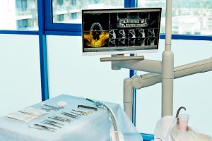
Digital pantomograph X-ray sFor making pictures of the facial skeleton in planning general stomatological and implant-prosthetic treatments. Thanks to this device we are able to classify a patient for a treatment and plan e.g. implantological treatment, as well as make repeated X-ray pictures in order to evaluate the effects of the treatment in time. This is of great importance in implantological treatment.

For making intraoral pictures, used in diagnostics and between procedures in root canal treatment, as well as in surgical-implantological treatment.
It allows to evaluate the state of dentition directly at the dental chair, and to visualise phases of treatments on the X-ray picture. Examples:
Has a tooth canal been filled properly; has a post been set precisely in a root; in the phase of making impressions on the implants and placing prosthetic crowns – have individual construction elements been properly screwed, set in the oral cavity.
Digital equipment, unlike analogue equipment, makes it possible to get pictures immediately after it has been taken and to visualise at the workstation, directly on a screen at the dental chair.
Visiting patients do not need to have the pictures taken at other X-ray laboratories.
An important asset of digital X-ray machines is that the dose of radiation is many times smaller than in analogue machines used before.
Stomatological microscope Microscope gives incomparably better possibilities for proper treatment, especially in root canal treatment.
 A considerable magnification of the visual field is reflected in considerably more precise draining, clearing, and filling tooth canals. It increases therapeutic possibilities, and therefore we are able to “save” teeth that had been classified for extraction before. We may treat complicated, seemingly hopeless cases, we may remove left, broken instruments from the canals, seal intracanal perforations, etc.
A microscope is also used in surgery of the alveolar process, e.g. cleaning inflammatory tissues in the bone, removing foreign bodies after injuries, surgical debribement of the alveolar process before implantation, regenerating bone structures.
The procedures are done more precisely and are less invasive for the patient.
Thanks to this apparatus we are able to carry out microsurgical procedures on the soft tissue in periodontal surgery (soft tissue transplants, plastic surgery of soft periodontal tissue and implants) and in procedures improving the aesthetics of prosthetic restorations.
Piezosurgery A device for cutting the bone tissue and for collecting bone transplants. It is now the most modern and sophisticated device used in alveolar process surgery and in implantology for making very precise incisions in the bone tissue with no harm to the soft tissue.

It is of great importance, especially when carrying out highly specialist bone transplant, sinus lift procedures, etc.
Except from the precision of the procedure, which means better prognosis in difficult cases, another merit is work safety. The Piezosurgery device eliminates the risk of damaging e.g. nerves or vessels passing through the maxillary bone structure and the mucous membrane of the maxillary sinus. The precision of the procedures is greatly improved, which considerably influences the effect of the treatment.